-
- (***) Towards Automated Semantic Segmentation in Prenatal Volumetric Ultrasound, TMI, 2019, paper
- (**) Medical breast ultrasound image segmentation by machine learning, Ultrasonics, 2019, paper
- (**) Cascaded Fully Convolutional Networks for automatic prenatal ultrasound image segmentation, ISBI, 2017, paper
- (**) Deep Attentive Features for Prostate Segmentation in 3D Transrectal Ultrasound, TMI, 2019, paper
- (***) Channel Attention Module with Multi-scale Grid Average Pooling for Breast Cancer Segmentation in an Ultrasound Image, IEEE transections on ultrasonics, 2020, paper
- (***) Auto-context Fully Convolutional Network for Levator Hiatus Segmentation in Ultirasound Images, ISBI 2018
Ultrasound Image Segmentation
(***) Towards Automated Semantic Segmentation in Prenatal Volumetric Ultrasound, TMI, 2019, paper
Problem
- the poor image quality, low contrast, boundary ambiguity and complex anatomy shapes conspire towards a great lack of efficient tools for the segmentation.
- It makes 3D ultrasound difficult to interpret and hinders the widespread of 3D ultrasound in obstetrics.
Contribuction
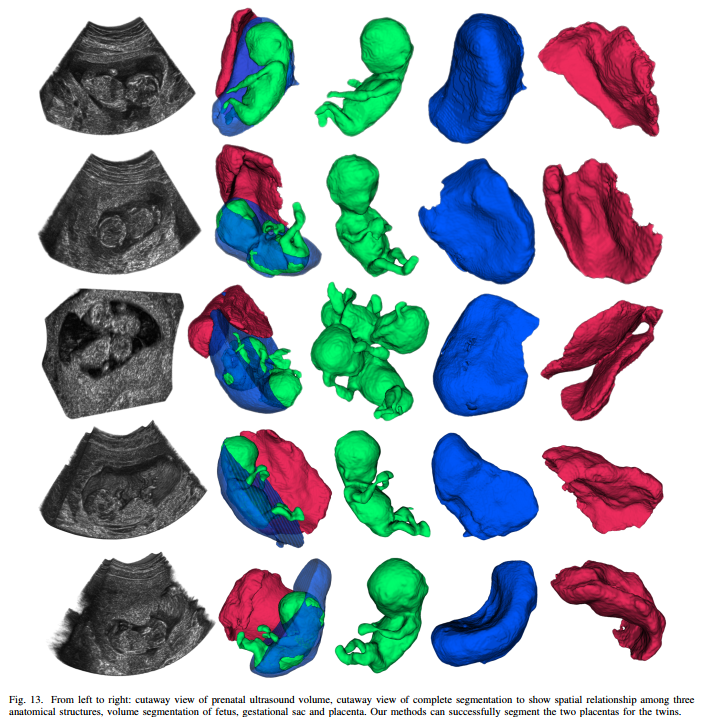
- we propose the first and fully automatic framework to simultaneously segment multiple anatomical structures with intensive clinical interest, including fetus, gestational sac and placenta, which remains a rarely-studied and arduous challenge
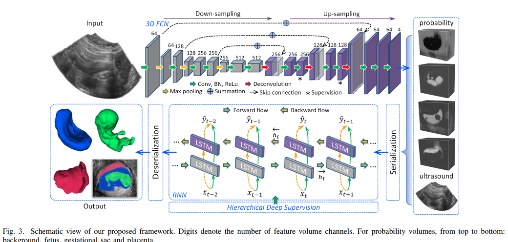
- we propose a composite architecture for dense labeling, in which a customized 3D fully convolutional network explores spatial intensity concurrency for initial labeling, while a multi-directional recurrent neural network (RNN) encodes spatial sequentiality to combat boundary ambiguity for significant refinement
- we introduce a hierarchical deep supervision mechanism to boost the information flow within RNN and fit the latent sequence hierarchy in fine scales, and further improve the segmentation results
Method
Result
Limitation and Discusion
- Currently, our framework consists of 3D FCN and RNN, which are two complementary but separately trained parts.
- However, currently, only ConvLSTM in 2D format has been reported for biomedical image segmentation
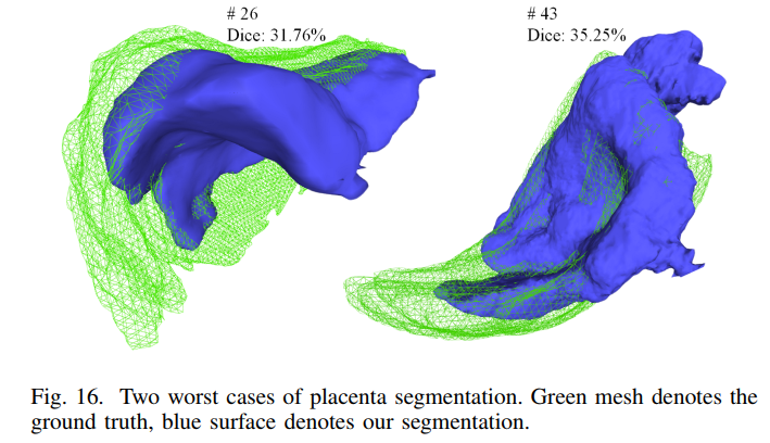
- High time efficiency is expected for our task. However, confined by GPU memory, we resort to the overlap-tiling stitching strategy for both FCN and RNN to generate the prediction of whole volume
- From the reproducibility experiments, we can see that different scanning directions and poses have impacts on the segmentation
(**) Medical breast ultrasound image segmentation by machine learning, Ultrasonics, 2019, paper
Problem
- manually segmenting the ultrasound images, which is skill and experience dependent, would lead to a subjective diagnosis
- it is time-consuming for radiologists to review hundreds of clinical images
Contribution
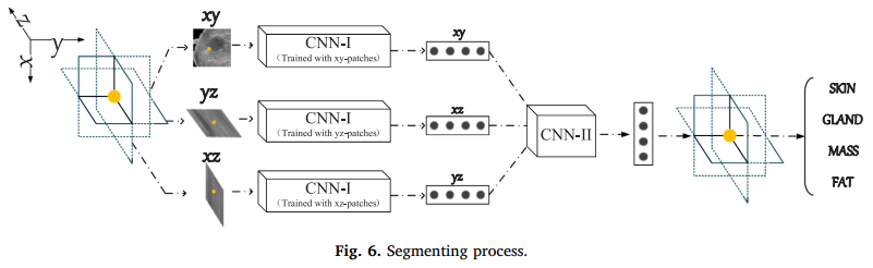
- In this paper, we propose to use convolutional neural networks (CNNs) for segmenting breast ultrasound images into four major tissues: skin, fibroglandular tissue, mass, and fatty tissue, on three-dimensional (3D) breast ultrasound images
Method
Result and Discusion
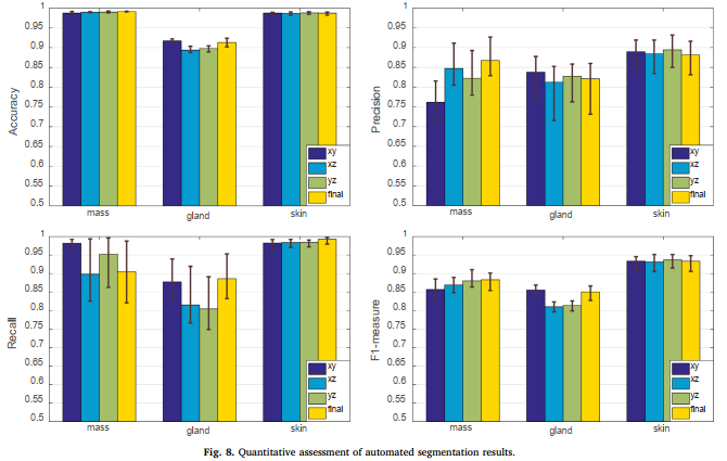
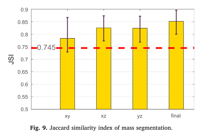
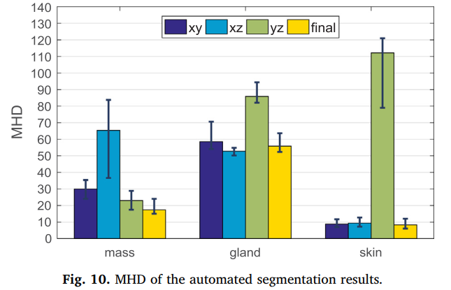
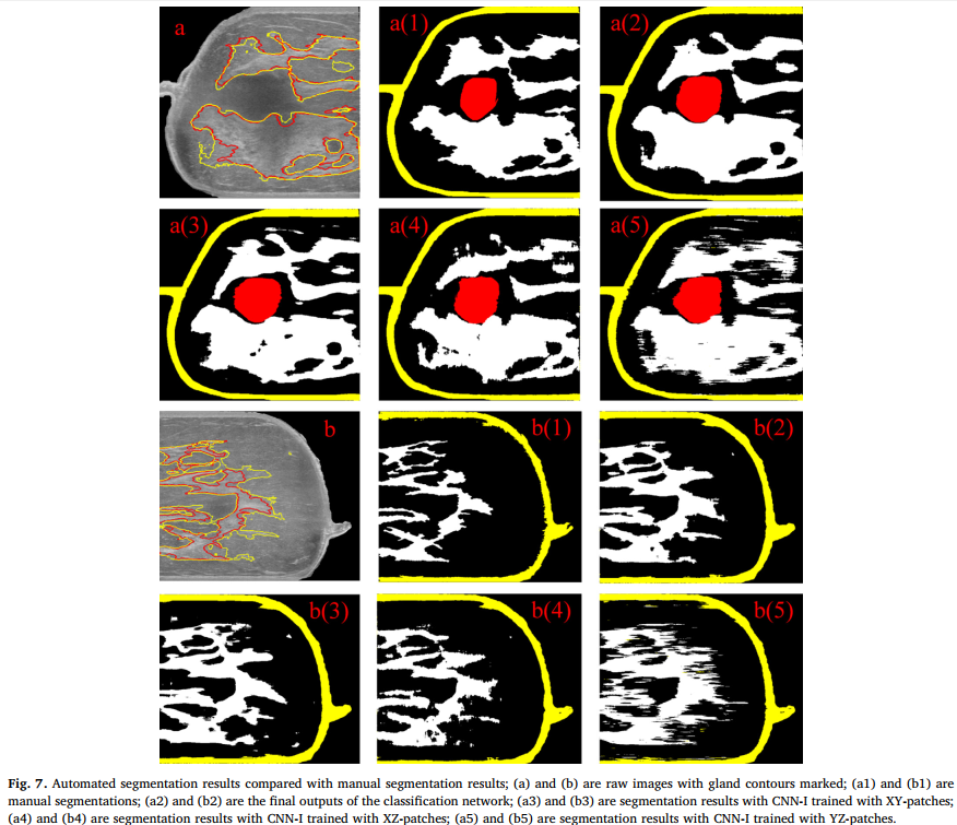
- the proposed automated segmentation method might have the potential to provide an objective reference for radiologists on breast image segmentation, so as to help breast cancer diagnosis and breast density assessments
(**) Cascaded Fully Convolutional Networks for automatic prenatal ultrasound image segmentation, ISBI, 2017, paper
pros and cons of ultrasound imaging
| pros | cons |
|---|---|
| 1. real time imaging 2. low cost 3. free of radiation | 1. various intensity distribuction 2. boundary ambiguities 3. structures deform due to different pressure |
Challenges
- Firstly, US images often present various intensity distributions due to different imaging conditions
- Secondly, a number of factors including acoustic shadows, speckle noise and low contrast between objects and surrounding tissues cause the typical boundary ambiguities and long-span occlusion
- Thirdly, different press sources tend to make the fetal anatomical structures deform.
Contribution
- We get a good starting point by tailoring the FCN-8s for considering both low model complexity and high segmentation accuracy
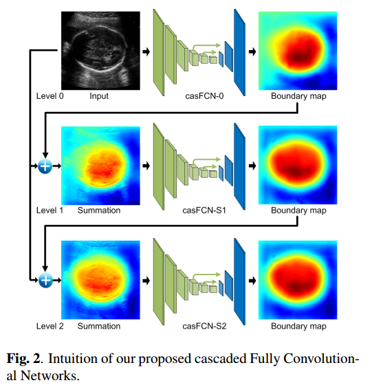
- Implanting our tailored FCN into an Auto-Context scheme enhances the boundary details by dynamically exploring local context, and thus gains remarkable improvement from level to level.
- Additionally, we provide a new perspective to the classical Auto-Context scheme with the investigation on two types of join operators.
Result and Conclusion
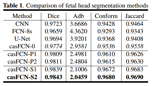
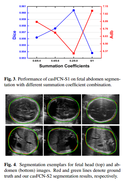
- The proposed casFCN framework is general and can be easily extended to other US image segmentation tasks.
(**) Deep Attentive Features for Prostate Segmentation in 3D Transrectal Ultrasound, TMI, 2019, paper
Problem
- manual outlining is tedious, time-consuming and often irreproducible, even for experienced physicians.
- the ambiguous boundary caused by poor contrast between the prostate and surrounding tissues
- missing boundary segments result from acoustic shadow and the presence of other structures (e.g. the urethra)
- inhomogeneous intensity distribution of the prostate tissue in TRUS images
- the large shape variations of different prostates
Contribuction
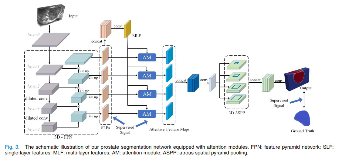
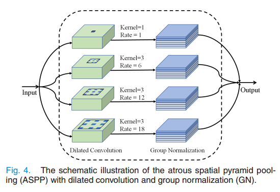
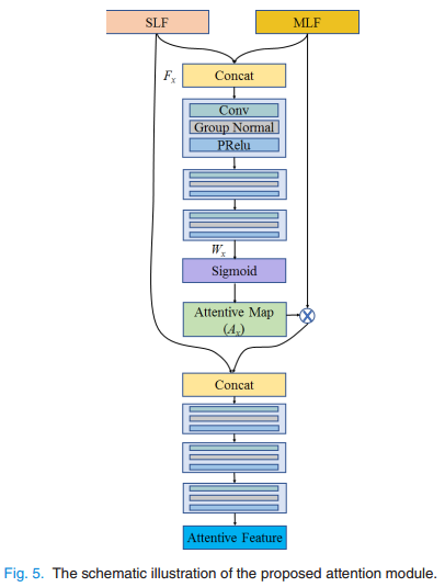
- We propose to fully exploit the useful complementary information encoded in the multi-level features to refine the features at each individual layer.
- We develop a 3D attention guided network with a novel scheme for TRUS prostate segmentation by harnessing the spatial contexts across deep and shallow layer
Result and Conclusion
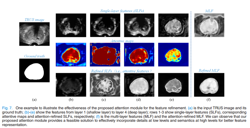
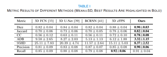
- Our key idea is to select the useful complementary information from the multi-level features to refine the features at each individual layer.
- Experiments on challenging TRUS volumes show that our segmentation using deep attentive features achieves satisfactory performance. In addition, the proposed attention mechanism is a general strategy to aggregate multi-level features and has the potential to be used for other medical image segmentation and detection tasks.
(***) Channel Attention Module with Multi-scale Grid Average Pooling for Breast Cancer Segmentation in an Ultrasound Image, IEEE transections on ultrasonics, 2020, paper
Problem
- recently proposed methods such as FCN, SegNet and U-Net still need to be further improved to provide better semantic segmentation when diagnosing breast cancer by ultrasound imaging, because of their low performance.
- only local information within receptive fields is used, global information on an input image is lost during the process of the architectures
Contribuction
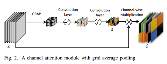
- we propose a novel channel attention module with multi-scale grid average pooling, that is capable of improving the performance of CNNs for breast cancer segmentation in an ultrasound image.
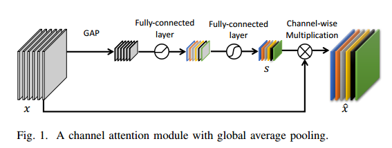
- we demonstrate that a channel attention module using grid average pooling instead of global average pooling can improve segmentation performance, by using the local information of an input image
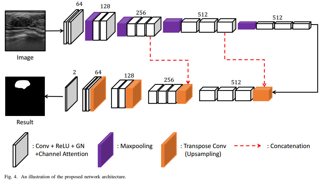
Result and Conclusion
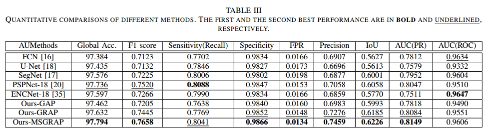
- The channel attention module with multi-scale grid average pooling (MSGRAP) allowed maintaining local and global information for the semantic segmentation of breast tumors in ultrasound images. In contrast, the channel attention module with global average pooling could only maintain global information
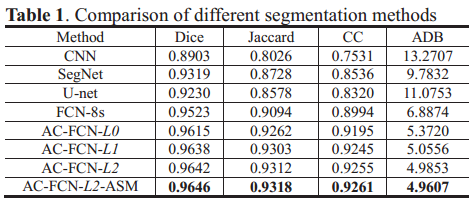
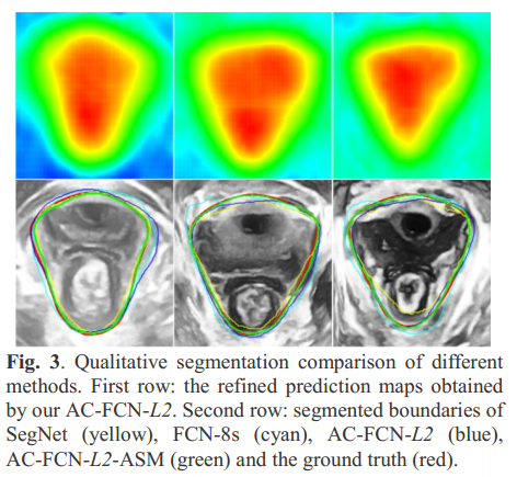
(***) Auto-context Fully Convolutional Network for Levator Hiatus Segmentation in Ultirasound Images, ISBI 2018
Problem
- Developing automatic methods for the levator hiatus (LH) segmentation is a necessary and crucial image-processing issue that needs to be resolved to achieve more objective and efficient female pelvic floor dysfunction (FPFD) examinations.
Contribuction
-
a customized FCN is integrated with the auto-context model for iteratively refining the prediction map.
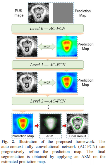
Result and Conclusion